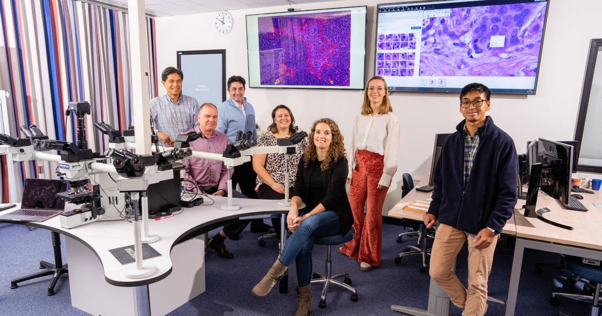
Research by UMC Utrecht shows that artificial intelligence can help pathologists evaluate the sentinel lymph node more efficiently. This evaluation is necessary to detect possible breast cancer metastases. Applying AI assistance to detect these metastases significantly reduces costs, is safe, saves pathologists time and makes their work more enjoyable.
The research group’s findings were published today in the Scientific Medical Journal Cancer of nature.
Read the English version >
“The demand for care and health care costs are increasing dramatically, and there is already a global shortage of pathologists,” says Carmen van Duijwaert, a clinical epidemiologist and postdoctoral doctor. “By using AI, pathologists can spend less time evaluating lymph nodes and we save costs (win-win), while the diagnosis remains safe for patients. We like this because it shows that if you apply AI in this safe way, you will not only save money and time It will also make the pathologists’ work more fun.
AI identifies metastases faster (and cheaper).
After a woman’s breast lump for breast cancer is removed, the sentinel node is often removed as well. This is the lymph node that first collects lymph fluid from the tumor. When breast cancer spreads, cancer cells first end up in the sentinel lymph node. This is why pathologists always carefully examine this tissue, because this can determine whether there are metastases. Since the implementation of digital pathology in 2015, it is no longer done using a microscope at UMC Utrecht, but behind a screen.
Pathologists sometimes see these metastases with the eye, but the smaller the metastases, the more difficult it is. Moreover, they have to check all the slides very carefully. To make sure no metastases are missed, pathologists examine sections (thin slices) of the glandular tissue again. They then use additional staining with antibodies that recognize tumor-specific proteins.
“Because these antibodies have a color tag, the cancer cells become clearly visible,” Carmen continues. “This way you can see if there are still cancer cells that were not detected during the first evaluation, without additional staining. These additional stains are (relatively) expensive, around €25 per tissue section, of which we already do (at least) five.” For each sentinel node. Sometimes multiple tissue blocks from multiple sentinel nodes must be examined, so costs add up very quickly and in addition, it is primarily an intensive and time-consuming task for the pathologist.
Faster and cheaper?
During the CONFIDENT-B trial, Carmen investigated whether artificial intelligence could help assess sentinel lymph node histology more efficiently. Could it be done faster, taking rising health care costs into account, and at a lower cost, without the risk of losing cancer cells if additional spots are deleted?
“You can actually use AI in two ways: completely autonomously (“Autonomous AI” – ed.) or as an assistant to a healthcare professional (“Assistant AI – ed.),” Carmen explains. “With autonomous AI, the algorithm decides completely independently whether there is a tumor or not. This is very exciting and currently unimaginable. What if the algorithm is wrong? This raises all kinds of ethical and legal questions: How safe is it to start using the algorithm in As soon as possible?
Carmen and her team actually used this other form of AI, which is AI assistance. The algorithm will help you then, but you still have to look at the result yourself. So the pathologist still looks at the sections.
“Our AI application, which we did not develop ourselves but licensed from Visiopharm, does the structure recognition,” says Carmen. “It “flags” whatever the pathologist thinks: ‘This doesn’t belong in a lymph node.’” The algorithm then puts a red, orange, or yellow circle around it, depending on how suspicious it is that the algorithm finds the spot.
These signs give pathologists a signal that they need to look at something, but they then do so in a much more targeted way and then don’t have to look at the entire section carefully.
Innovation that reduces health care costs
During the experiment, Carmen worked with two sets of sentinel nodes. For one group, the standard procedure was followed, and for the other group, AI was used first. In the AI group, Carmen first ran the AI application, and then the pathologists looked specifically at the circled spots. This examination is performed quickly by the pathologist, often without the need for additional expensive staining.
And imagine what? Less time was needed in the AI group, and it turned out to be much cheaper to work this way. “During the trial, we have already saved 3,000 euros in additional coloring. This can amount to tens of thousands of euros per year,” says Carmen.
The amount saved only increases when you take into account that in some hospitals, expensive additional staining is often started immediately in order to work faster. The paper also develops several scenarios that will allow other pathology laboratories to calculate the potential cost savings of using AI for this application.
Naturally, an additional check was performed during the trial to determine whether the AI procedure was safe: in all cases where no tumor cells were detected, additional staining was performed, and the AI was found to be correct. In addition, the unanimous conclusion of the participating pathologists was that working with the algorithm makes their work more enjoyable.
Paul van Diest, Head of Pathology and Head of Research: “This research has shown that using AI we can detect lymph node metastases safely, quickly and cheaply. This makes a significant contribution to the commercial viability of introducing AI into pathology, because unfortunately there is no compensation for this yet.”
Pathologists at UMC Utrecht now work with this method as a standard for breast cancer, and perhaps the first of its kind in the world.
Questions, comments or tips for editors?

“Total coffee specialist. Hardcore reader. Incurable music scholar. Web guru. Freelance troublemaker. Problem solver. Travel trailblazer.”


More Stories
GALA lacks a chapter on e-health
Weird beer can taste really good.
Planets contain much more water than previously thought