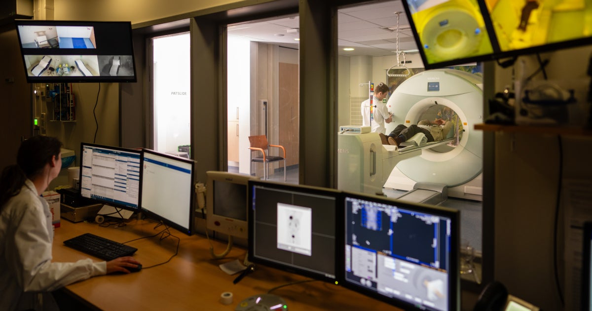
How can artificial intelligence be used to better treat cancer patients? Researchers from UMC Utrecht will answer this question. Four research projects at UMC Utrecht have received funding from the Hanarth Fund.
Read in English >
The Hanarth Fund grew out of the legacy of Arthur del Prado, founder and former CEO of ASM International. The fund focuses on publishing artificial intelligence (Amnesty International) and Machine learning To improve the diagnosis, treatment and outcomes of cancer patients. More than 6 million euros have been awarded to various projects this year. Read more about the different projects awarded at UMC Utrecht below.
Rapid identification of tumor type
MaLMec: Machine-Learned DNA Methylation Classification to Enable Tumor Subclassification of Liquid Biopsies
Lead applicant Jeroen de Ridder's (Associate Professor) project focuses on tumors of the central nervous system. Reaching the tumor is often impossible without surgery. “This poses a challenge for neurosurgeons,” Jeroen says. They have to perform surgery on a tumor without knowing the type of tumor. This means that there is a possibility that the patient may need further surgery. “Sturgeon” has recently been developed for this purpose. This is an application of artificial intelligence through which the type of tumor can be accurately determined during the operation. This allows the appropriate surgical strategy to be applied immediately. Jeroen and his team will focus on developing a different type of sturgeon. With this new alternative it is sufficient to use cerebrospinal fluid instead of (part of) the tumor. This will make it possible to determine the type of tumor even before the operation. The main challenge is that the cerebrospinal fluid contains a mixture of tumor and normal DNA. Artificial intelligence models will be trained to take this into account.
Evaluation of the effect of radiotherapy (chemotherapy).
Improving detection of residual disease after radiotherapy (chemotherapy) for locally advanced head and neck squamous cell carcinoma
Lead applicant Nico van den Berg (Professor of Computational Imaging) and his team are focusing on head and neck cancer in their new project. Objective: To develop a clinical artificial intelligence model for radiologists and radiotherapists. In head and neck cancer, correctly assessing the effect of radiotherapy (chemo) is a major challenge. The form will make this easier. “The goal is to reliably distinguish between residual disease and changes after treatment,” Nico says. “This will allow a more accurate assessment of whether a second operation is needed. This could improve survival and quality of life. Unnecessary procedures are reduced to a minimum and the burden on the patient is reduced.
Identifying prostate cancer metastases
Towards individualized PSMA PET/CT-guided therapy in metastatic prostate cancer using machine learning-derived risk stratification (custom-designed)
Associate Professor Arthur Pratt's project focuses on metastatic hormone-sensitive prostate cancer. Good detection of metastases is essential for choosing the appropriate treatment. Nowadays, an improved technique for metastasis detection (PSMA PET/CT) is used in daily clinic. However, the way to classify the extent of metastases for treatment selection is to use an older technique (bone scan). The new classification is necessary to determine the appropriate treatment at this time. We can also gain more knowledge about differences between patients through new scientific studies. “In this project, we will apply artificial intelligence (AI) to automatically analyze PSMA PET/CT,” explains Arthur. “This gives us more and better insight into the differences between patients.”
Better MRI
Physics-informed neural networks for unifying brain MRI: enhancing AI applications in gliomas and meningiomas.
The project by Associate Professor Alessandro Sprezi and Assistant Professor Stefano Mandiga focuses on a rare brain cancer. Thanks to artificial intelligence tools and MRI scans, the size and location of these tumors can be better estimated. However, these AI tools are not yet widely applicable. “We will expand the training sets on MRI images to enhance the AI tools,” say Alessandro and Stefano. Mainly in the field of rare brain cancers such as meningiomas and gliomas. There are currently no large standardized datasets available for this purpose. This will also make these AI tools applicable to rare brain cancer.
Friends of UMC Utrecht and Wilhelmina Children's Hospital
These studies can begin thanks to the support of the Hanarth Fund of the Friends UMC Utrecht & Wilhemina Children's Hospital, the charitable foundation of the (children's) hospital. Read more About how research into a new treatment is being developed at UMC Utrecht.
Questions, comments or tips for editors?

“Total coffee specialist. Hardcore reader. Incurable music scholar. Web guru. Freelance troublemaker. Problem solver. Travel trailblazer.”


More Stories
GALA lacks a chapter on e-health
Weird beer can taste really good.
Planets contain much more water than previously thought